Anatomy of the Eye
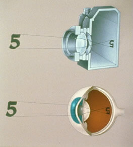
The eye is a complex organ that works much like a camera, focusing light rays and forming an image. The surface of the eye is the cornea, a thin layer of tissue that provides a clear window for light to pass through. The cornea bends or refracts light rays so they focus precisely on the retina. Next is the iris, the colored part of the eye. In the center of the iris is the pupil. The iris functions like a shutter, adjusting pupil size to control the amount of light entering the eye. Located behind the iris is the lens, which works together with the cornea to focus light. Like the lens in a camera, the lens of the eye adjusts light rays as vision shifts between near and far objects. Light then passes through the vitreous, the gelatinous substance that fills most of the eye. The back of the eye is lined with a thin layer of tissue containing millions of photoreceptor (light-sensitive) cells. This is the retina, where light rays focus. The retina is like the film of a camera.
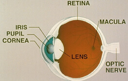
The center of the retina is called the macula. The macula is responsible for clear central vision and the peripheral retina is responsible for peripheral vision. The retina converts the image into an electrical signal that travels down the optic nerve to the brain.
Retinal Exam and Testing
Retinal Exam
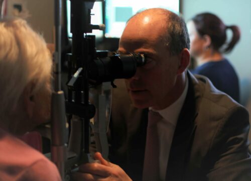
In order to examine the retina, first it is necessary to obtain a full medical and ocular history. Afterward, the patient’s vision is checked using the eye chart. The pupils are examined. The eye pressure is taken, then the eye is dilated with short-acting, pupil-dilating eye drops. Dilation of the pupils is necessary for the doctor to examine the entire anatomy of the inner part of the eye. At this point, the patient is brought into the exam room and the physician begins his examination.
The exam includes a complete ophthalmic assessment from the front of the eye to the back of the eye. The retinal exam includes a full inspection of the vitreous cavity and examination of the optic nerve, macula, retinal blood vessels and extreme peripheral retina. Once this is done, your doctor will make the decision whether or not to perform various retinal tests to further clarify the vitreous, macula, retina or choroidal diagnosis.
Retinal Testing
Dr. Barofsky can perform many retinal tests in order to confirm the diagnosis.
OCT Scans
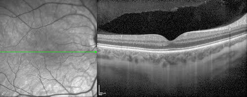
Optical Coherent Tomography (OCT) is a method used to image the retina and quantitatively track changes that occur. OCT analysis takes only seconds to perform and uses advanced optics and physics in order to measure retinal thickness, volume and area. Subtle retinal defects can easily be seen by your doctor.
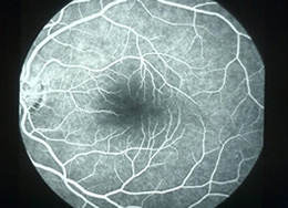
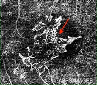
Fluorescein Angiography (FA)
Fluorescein angiography is a diagnostic test used to evaluate the retina and the retinal blood vessels with the help of a contrast dye (fluorescein). These pictures help the doctor evaluate the retina and diagnose and track problems such as diabetic retinopathy, macular degeneration, abnormal blood vessel growth, retinal swelling, retinal detachment, cancer and retinal tumors.
First, the patient’s pupils are dilated with eye drops. Then a few photographs are taken with a special ophthalmic camera. Next, the contrast dye is injected, usually in the patient’s arm. The dye travels up to the eye within a few seconds and “lights up” the retinal blood vessels and retinal structures. This test only takes a few minutes to perform.
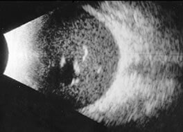
Ophthalmic Ultrasound
Dr. Barofsky can perform an ophthalmic ultrasound using simple sound waves that reflect off different tissues in the eye to form an image.
This test is used to determine the extent of a retinal detachment, evaluate the retina and vitreous behind an advanced cataract or hemorrhage and determine the thickness of an intraocular tumor. This test takes seconds to perform.
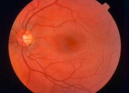
Retinal Photography
Photographs can be taken of the retina to track changes over time. Also, lesion size can be determined using special software.
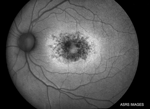
Fundus Autofluorescence
A new technique called fundus autofluorescence (FAF) allows for imaging of the retina and identification of retinal diseases that might not be readily apparent. It can determine metabolic changes within the deep retina that are not able to be seen during a routine examination. This approach can be especially helpful to analyze patients with vision loss of undetermined origins or those with a family history of hereditary retinal diseases.

Indocyanine Green Angiography (ICG)
This is another excellent method for visualizing retinal circulation. In contrast to fluorescein angiography, ICG is of particular value in studying choroidal circulation while investigating macular disease.
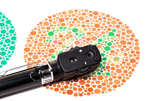
Color Vision Testing
Color vision tests are important to determine the diagnosis of various retinal diseases.
OCT Angiography
Is a non-invasive imaging technique used to visualize retinal blood vessels in 3-D. This ultra-high-resolution image can help with diagnosis and treatment of age-related macular degeneration, diabetic retinopathy, retinal artery occlusion, and retinal vein occlusion.

Educational Links
- American Academy of Ophthalmology
- American Society of Retinal Specialists
- Prevent Blindness America
- eyeSmart® – Macular Degeneration and Low Vision
- The Ocular Immunology and Uveitis Foundation – Education
- MD Support – The Eyes of the Macular Degeneration Community
- American Diabetes Association
- American Macular Degeneration Foundation
- National Eye Institute
- EyeWiki™
- Foundation Fighting Blindness®
- National Eye Institute – Macular Hole
- National Eye Institute – Macular Pucker
- National Eye Institute – Retinal Detachment
- eyeSmart® – Eye Health Information for the Public from the American Academy of Ophthalmology
- ASRS – Retina Health Information and Fact Sheets

