
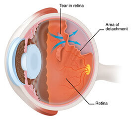
The retina is a photosensitive tissue within the eye that allows us to see. The retina lines the back of the eye similarly to how camera film lines the back of a camera.
Rhegmatogenous Retinal Detachment – This is the most common type of retinal detachment, which occurs when retinal tears are formed and vitreous fluid leaks under the retina causing a separation of the retina from the back wall of the eye. Without treatment this will lead to blindness.
Risk factors for retinal detachment:
- Retinal tear
- Posterior vitreous detachment
- Myopia (near sightedness)
- Injuries to the eye
Retinal detachment is an ocular emergency that requires immediate treatment. Symptoms would include:
- Sudden onset of floaters
- Flashing lights
- A shadow or curtain coming from the side of the visual field toward the center
- Loss of peripheral vision
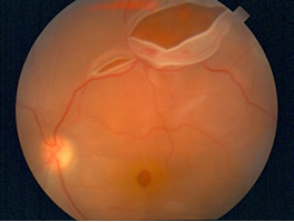
Diagnostic Testing
The doctor performs a full dilated ophthalmic and retinal examination to determine the type and extent of retinal detachment.
Treatments
- Laser photocoagulation – Small and early retinal detachments can often be treated with in-office laser photocoagulation alone.
- More extensive retinal detachments can be treated with various methods such as:
- In office pneumatic retinopexy
- Pars plana vitrectomy with gas injection
- Pars plana vitrectomy with scleral buckle and gas injection
- Scleral buckle alone


